Laboratory tests for the diagnosis of Infectious Bronchitis (IB)
Laboratory tests that can be done for a definitive diagnosis of IB are:
Virus Isolation – Antigen detection
Virus isolation is usually done in 9–10 day of age embryonated specific pathogen free (SPF) eggs. Several blind passages may be necessary before clinical signs characteristic of IBV are observed in embryos. Typical lesions in embryos occurring at about 5–7 days post inoculation are curling and dwarfing of the embryos, clubbing of down, red or haemorrhagic embryos, and possibly white urate deposits in kidneys. Tracheal organ cultures (tracheal rings) may also be used for isolation of IB viruses. In this case, ciliostasis and damage to the tracheal epithelium are seen within 48 to 72 hours of inoculation, when the tracheal rings are observed under low power microscopy. This method gives results more quickly than does the use of embryonated eggs, but the identity of the isolate as IB must be confirmed by other methods since IBV is not the only pathogen that may cause ciliostasis of tracheal epithelium. See the ciliostasis test.
Immunofluoresence Test with fluorescein-conjugated antibodies that attach to the IBV when present in tracheal smears.

Virus Isolation-genome detection
- RT–PCR – is used with specific primers to detect the genome of the virus in tracheal samples or inoculated embryonated eggs or tracheal organ cultures.
- RFLP, – which recognizes the genome of the IBV and is used to determine the genotype.
- IBV typing by nucleic acid sequencing.
- IBV typing by real–time RT–PCR.
Antibody determination
Testing serum samples at intervals (for example at the time of the clinical signs and 2 or 3 weeks later) provides the basis for serological diagnosis. This is also applicable for monitoring vaccination results.
Agar Gel Precipitation Test (AGP)
A short time after infection a sudden rise in precipitating antibodies occurs, followed by a decline thereafter. The test is not serotype specific but can be useful to detect a recent IB infection. Although it has the advantage of being quick and easy to perform, it is very insensitive and would only be used as a quick way to detect a recent infection. For this test, two holes are punched in an agar gel and known IBV antigen and sera from suspect birds are allowed to migrate through the gel. An interaction between the antigen and any antibodies specific for IBV in the gel, results in precipitation of the antigen, which shows up as a visible line of identity in the gel.
Virus Neutralisation test (VN)
The virus neutralisation (VN) test is by far the most accurate method available for differentiating between IBV serotypes as well as for confirming the identity of new ones. To perform the test it is necessary to have a culture of each IB virus of interest as well as monospecific antiserum to each one. These specific antisera are very important if accurate differentiation of the serotypes is to be achieved. Each one is prepared by inoculating intranasally a group of SPF chickens with one of the IBV serotypes of interest. Blood is collected from the chickens 3 to 4 weeks later and the serum obtained from this blood contains serotype specific antibodies to the IB virus inoculated.
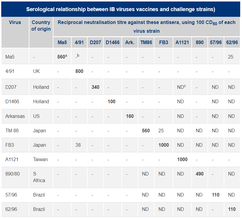
In a laboratory assay system, such as embryonated chicken eggs or tracheal organ cultures, each virus of interest is tested against the specific antisera raised to each of these IBVs. This can be done in two ways; by testing one dilution of each antiserum against a series of virus dilutions, or (more accurately) by testing a known amount of each virus against a series of dilutions of the antiserum. In this way, as shown below, it is possible to produce a Table showing the titre of each IB virus against the serum to the homologous (the same) IBV serotype as well as all the heterologous (different) IBVs. The higher the titre, the greater the relationship between the IBVs. Thus it is possible to say whether a field isolate is a “new” variant or is related to a known IBV serotype.

aHomologous titre in bold
bTitre </= 1:20
cNot done
Haemagglutination Inhibition Test (HI)
IBV does not spontaneously agglutinate chicken red blood cells, therefore it needs to be treated with the enzyme neuraminidase before it can be used in the HI test.
The test is a possible alternative to the VN test, as it is much simpler and quicker. The HI test may be serotype specific, but only if it is performed by experienced workers, under tightly controlled laboratory conditions. This is because, when chickens have been exposed to more than one different IB serotype, there is a higher level of cross reactions between IB serotypes in this test than in the VN test.
Enzymelinked immunosorbent Assay (ELISA)
Specific antibodies in serum bind to the virus, which is attached to the bottom of a 96–well plastic plate. The complex is detected by an enzyme–labelled anti–γ-globulin. After adding an enzyme substrate, a resulting coloured product is measured. This assay is not serotype specific, but is useful as a flock test to confirm adequate antibody responses to, for example, vaccination, or to give an indication of a recent or current IB infection. Commercial kits are available.
